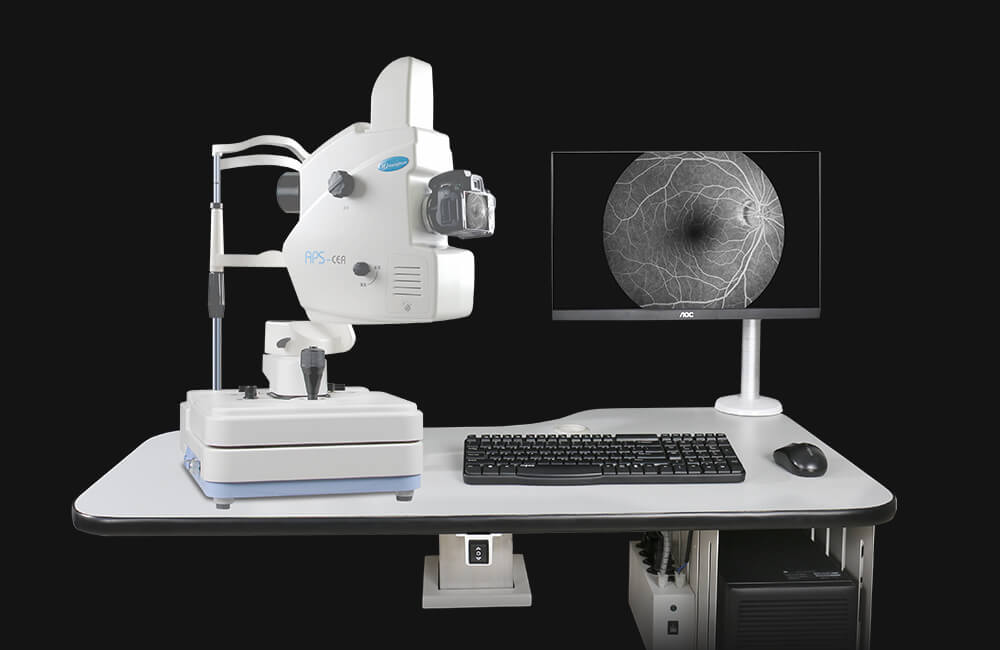
Digital Fluorescein Angiography and Fundus Photography
There are a few components that make up the angiography and fundus photos system. These are: A large camera, computer server, and a color monitor. A trained retinal photographer takes high resolution photographs of the patient’s retina following which the pictures are immediately accessible for review by the eye doctor.
Digital retinal photography uses high resolution photographs of the retina and macula by using large cameras. The test is performed, with the patient seated in the chair. Digital retinal photography has numerous clinical usages. For instance, it can be used for precise documentation of the extent of retinal disease at one point at a time, so that it can be compared at a later date.
Fluorescein (a yellow synthetic dye) angiography is used to take additional pictures with dye contrast in some retinal diseases. Fluorescein is injected into the vein in the arm. This circulates rapidly into the eye. Fluorescein will highlight the blood vessels and most areas of retinal disease when the camera is used with certain light filters. This helps with assessing severity and extent of retinal diseases which would help with clinical decision making. For instance, fluorescein angiography is usually utilized to assess age-related degeneration, diabetic retinopathy, and retinal vascular occlusions.
In the past, 35 mm films were used for retinal photography and angiography. Because these films had to be developed it would cause some delays. With advance technology, digital angiography and photography offer rapid and precise results which are viewed on a computer monitor.