The Retinal Examination
When you visit a retina specialist then he would use instruments and special lenses to directly visualize the retina.The most important part of the evaluation process is the retinal examination. Primary eye doctors use the same tools as employed by retina specialists. Most patients are already familiar with these instruments.
At the time of the retinal examination, eye drops are used to dilate the pupils. Dilating the pupils allows the doctor to look at both the central and peripheral retina thoroughly. The pupils might remain dilated for several hours after the examination. Most patients are ok to drive home after dilation of pupil. If you’ve never been dilated before or if you have had blurry vision in the past after receiving dilating drops, then we suggest that you bring someone with you to drive you home safely.
The retina specialist use two instruments to look at the retina. These are the slit lamp and the indirect ophthalmoscope. The retina itself is often visualized directly by using special lenses and lights. The lights used during the retinal examination are not harmful for the patient’s eye. If abnormalities are identified during the retinal examination then further diagnostic testing may also be required for a more detailed and precise evaluation.
When you visit a retina specialist then he would use instruments and special lenses to directly visualize the retina.The most important part of the evaluation process is the retinal examination. Primary eye doctors use the same tools as deployed by retina specialists. Most patients are already familiar with these instruments. There are many retina surgeons in Houston area and most retina doctors in Houston would have similar examination techniques.
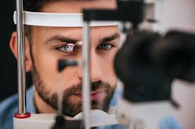
Slit-lamp Examination:
The Slit-lamp is a commonly used tool to examine the retina. A bright light is utilized to view the inside of the eye with the patient seated upright. By using the magnifying lens, eye specialists utilize the slit-lamp to examine the macula and optic nerve in great detail.
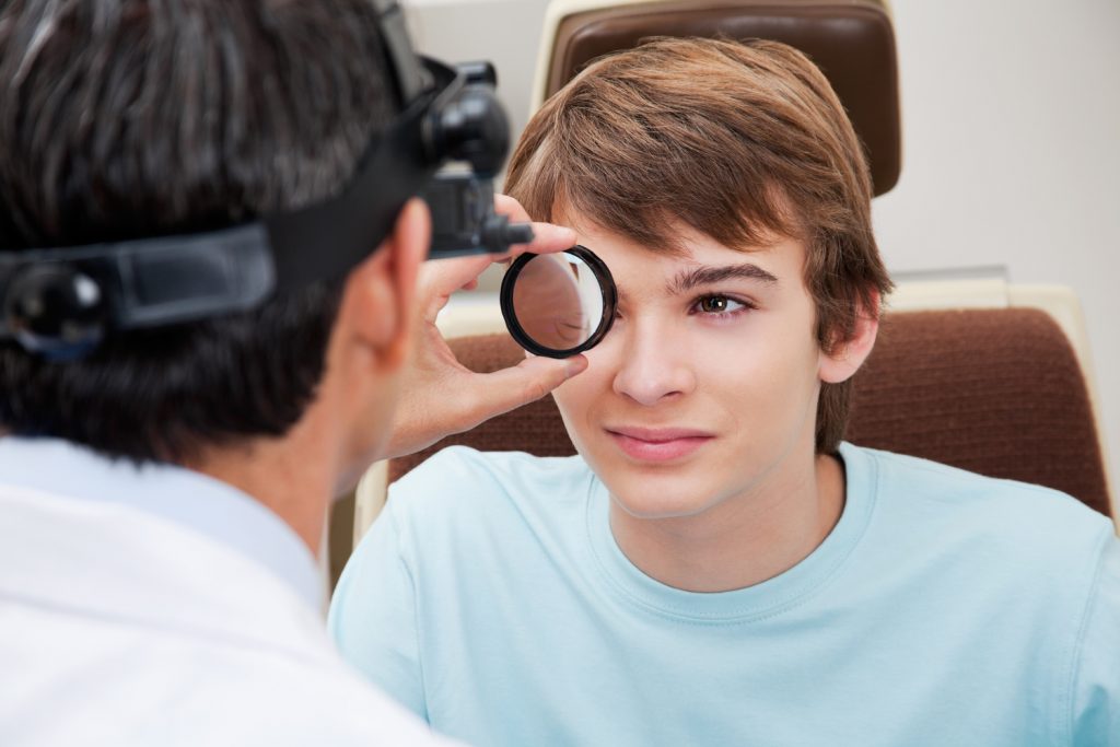
Indirect Examination:
Another magnifying tool to examine the peripheral retina is the indirect ophthalmoscope. The patient’s eyes are examined while the patient is sitting upright or lying back. The doctors wear a headpiece consisting of unique mirrors, lenses, and a light source. This allows the doctor to look at different parts of the peripheral retina comprehensively with the help of a handheld lens. Occasionally, the indirect examination is mixed with a maneuver known as sclera indentation. This involves putting mild pressure on the eyelids to look at the peripheral retina.